Dental Aesthetics
As well, if the patient has some other concerns or questions about the aesthetics added to solving the problems of dental health we help in any decisions by explaining in detail de pros and cons of each step in the treatment.
SOLUTIONS in mild cases. Minimally Invasive Dentistry.
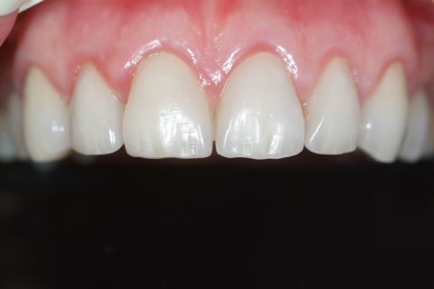
If the front teeth area has been minimally affected the dental surface can be rehabilitated by porcelain veneers or laminates (truly porcelain nails) that have a minimal thickness and are fully made in the dental laboratory. And with them we obtain the best aesthetics results in modern odontology.
-
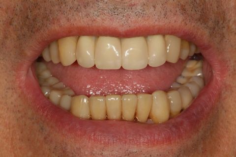
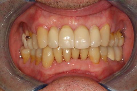
Old feldspathic veneers and a metal-porcelain crown
-
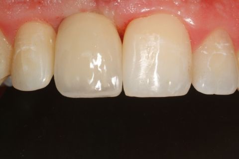
New feldspathic veneers and a e.max lithiumdisilicate crown
DENTAL SOLUTIONS FOR MAJOR DAMAGE. CAD-CAM TECHNOLOGY.
To solve more advanced or serious dental injuries (partial or complete crowns) and within the umbrella of aesthetic protheses we offer the patient the option to reconstruct those teeth with the latest in CAD-CAM technology (computer assisted design - computer assisted manufacturing) the most innovative technique in fixed prothesis and odontological rehabilitations, extremely precise and of the highest quality associated with maximum resistance and an important advance in conventional odontology.
CAD-CAM TECHNOLOGY IN THE TEETH
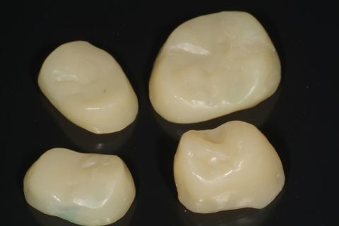
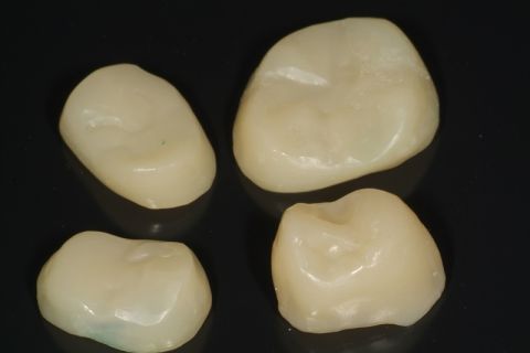
Partial or complete crowns of ceramic made of lithium disilicates IPS e.max of Ivoclar Vivadent.
-
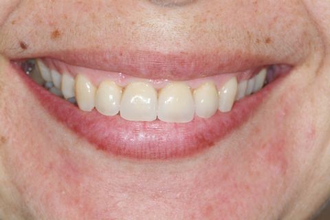
Old metal-ceramic crowns
-
e.max lithiumdisilicate crowns
-
Upper front teeth with multiple fillings
-
e.max lithium disilicate crowns
-
Four worn teeth
-
Four feldspathic veneers
-
Four feldspathic veneers
-
Oral situation before treatment
-
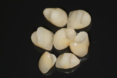
Seven e.max crowns
-
View of the inside of the e.max crowns
-
Oral situation after treatment
-
Previous deconstructed front
-
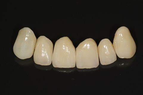
Six e.max crowns
-
View of the inside of the e.max crowns
-
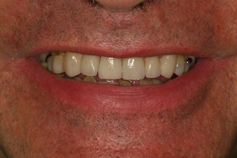
The six e.max crowns placed
-
End view of smiling
-
 Upper e.max crowns
 -
 Premolar and molar e.max crowns
 -
 Upper frontal e.max crowns
 -
 Right side view
 -
 Left side view
 -
 General view
CAD-CAM technology in teeth darkened by tetracyclines
The new crowns replace old teeth veneers that where covering teeth darkned by tetracyclines. The explanation of the darkening of the teeth is as follows: Because of infectious problems some antibiotics called tetracyclines where administered to young children. These tetracyclines bind to circulating calcium in the blood and accompany it to the teeth and bones. As tetracyclines have a dark color, teeth get stained irreversibly. Today only tetracyclines are not administered to children.
-
Tetracycline stained teeth and treated once with feldspathic venners
-
Teeth with modern crowns
-
Tetracycline stained teeth
-
Teeth with modern crowns
CAD-CAM technology in large composite restorations
Large restorations of composite Lava Ultimate of 3M.
-
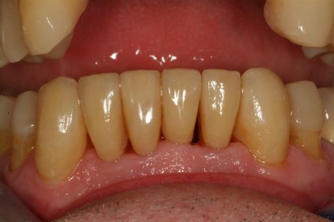
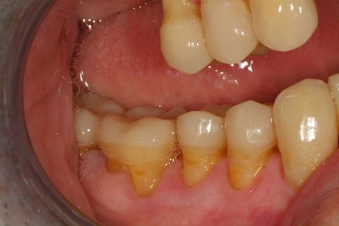
 The teeth are badly eroded
 -
 The teeth are retouched to receive crowns LAVA
 -
Complete LAVA overlays
-
The case concluded
-
The case concluded
CAD-CAM TECHNOLOGY ON IMPLANTS
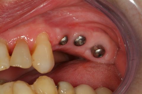
Once placed the implants in the bone, we wait a reasonable time and then we placed the teeth using CAD-CAM technology. Metallic structures are manufacturated and porcelain or acrylic teeth are added on them. In our clinic we receive CAD-CAM structures to the manufacturer of our own implants (BTI in Vitoria, See bti-biotechnologyinstitute.com) and in other cases the company Createch Medical (see Link createchmedical.com) based in Mendaro, both leading researches in metalworking and precision.
-
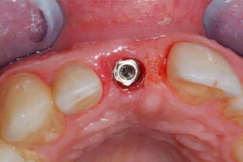
Implant placed
-
View of separated stump and crown
-
View of fitted stump and crown
-
View of fitted stump and crown
-
Implant in its place
-
The stump with porcelain
-
The stump with porcelain
-
Front view of the crown on the implant
-
Rear view of the crown on the implant
-
Two implants
-
Two pieces bridge on two implants
-
Three implants
-
Three pieces bridge on three implants
-
Front view of porcelain teeth on multiple implants
-
Right side view of porcelain teeth on multiple implants
-
Left side view of porcelain teeth on multiple implants
-
Panoramic radiograph with all the implants
Bridge
A bridge is the replacement of a tooth or several teeth carving the neighbours whom we shall call pillars, thus creating a block of all the pieces involved which may be from three to all the upper or lower denture.
They are structures of ceramic and metal or only ceramic in short spaces. They are fitted if the mouth does not possess a solid bone base to accept an implant (nowadays made very infrequent by the use of bone and gum grafts) and besides we must carve teeth that may be intact.
-
A metal-porcelain bridge of three pieces
-
Placed in the mouth
General Rehabilitation combining previous treatments.
-
 Veneers, bridges on implants and metal-porcelain crowns
 -
 Veneers, bridges on implants and metal-porcelain crowns
-
Disorganized mouth with placed implants and poor hygiene
-
Case concluded with crowns on implants, e.max crowns and LAVA Ultimate composite
-
Old incorrectly adjusted and fractured crowns and bridges
-
Rehabilitation with e.max crowns, feldspathic veneers, metal-ceramic bridges and bridges on implants
-
Before
-
Situation of the upper teeth before
-
Situation of the lower teeth before
-
Prostheses for the rehabilitation of the lower teeth
-
e.max crowns for upper right teeth
-
e.max placed in the mouth
-
LAVA Ultimate for molars and premolars
-
LAVA Ultimate and e.max on teeth
-
e.max placed on the lower anterior teeth
-
LAVA Ultimate placed in lower right premolars and molars
-
LAVA Ultimate placed in lower right premolars and molars
-
Metal-porcelain bridge in lower left posterior teeth
-
Left side in occlusion
-
Right side in occlusion
-
Upper skeletal
-
Upper skeletal
-
The end, with the upper skeletal placed in closed mouth
-
Open mouth
-
Before
-
After, solved case


